How Heart Health Impacts Overall Wellness: Why ECG Matters
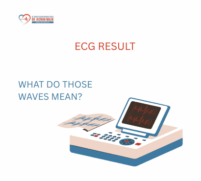
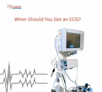
The heart is one of the most vital organs in the human body. It works continuously to pump blood, delivering oxygen and nutrients to tissues while removing waste products. Because the heart plays such a central role in circulation, its health has a direct impact on the functioning of every other organ system. When the heart is functioning properly, the body receives the oxygen and nutrients needed to maintain energy, immunity, and organ performance. However, when heart health is compromised, it can lead to a wide range of problems that affect overall wellness. Conditions such as irregular heart rhythms, blocked arteries, or heart muscle damage can disrupt circulation and cause serious health complications if not detected early. One of the most important diagnostic tools used to evaluate heart health is the electrocardiogram, commonly known as an ECG. This simple and non-invasive test records the electrical activity of the heart and helps detect abnormalities in heart rhythm, structure, or function. Understanding how heart health influences overall well-being and why ECG testing is important can help individuals take proactive steps to protect their cardiovascular health. This article explores the connection between heart health and general wellness, explains how ECG works, and highlights why regular heart monitoring is essential. Understanding Heart Health Heart health refers to the proper functioning of the heart and blood vessels. A healthy heart pumps blood efficiently, maintaining stable circulation throughout the body. The cardiovascular system includes: The heart Blood vessels such as arteries and veins Blood that carries oxygen and nutrients Together, these components support essential bodily functions including brain activity, muscle movement, digestion, and immune response. When the cardiovascular system functions properly, the body operates efficiently. However, disruptions in heart function can lead to widespread health issues. Why Heart Health Affects the Entire Body Because the heart pumps blood to every organ, problems with the heart can affect multiple systems simultaneously. Brain Function The brain requires a constant supply of oxygen-rich blood to function properly. Reduced blood flow can lead to dizziness, confusion, or even stroke in severe cases. Kidney Health The kidneys depend on healthy blood circulation to filter waste products from the body. Poor heart function can impair kidney performance. Energy Levels When the heart struggles to pump blood efficiently, the body may not receive enough oxygen. This can result in fatigue and reduced physical endurance. Immune System Healthy circulation helps transport immune cells throughout the body to fight infections. This interconnected relationship explains why cardiovascular health plays such a major role in overall wellness. Common Heart Problems That Affect Wellness Several heart-related conditions can impact general health. Coronary Artery Disease This condition occurs when the arteries supplying blood to the heart become narrowed or blocked. Reduced blood flow to the heart muscle may cause chest discomfort, fatigue, or heart attacks. Heart Rhythm Disorders Irregular heart rhythms, known as arrhythmias, can cause the heart to beat too fast, too slow, or irregularly. Symptoms may include palpitations, dizziness, or fainting. Heart Failure Heart failure occurs when the heart cannot pump blood effectively to meet the body’s needs. This condition can cause fluid buildup, shortness of breath, and fatigue. High Blood Pressure High blood pressure increases strain on the heart and blood vessels, raising the risk of heart disease and stroke. Early detection and management are essential for preventing complications. What Is an ECG? An electrocardiogram (ECG) is a medical test that records the electrical signals generated by the heart as it beats. Every heartbeat begins with an electrical impulse that travels through the heart muscle. These signals coordinate the contraction and relaxation of heart chambers, ensuring efficient blood circulation. An ECG measures these electrical signals and displays them as waves on a monitor or printed graph. Doctors analyze these patterns to evaluate heart rhythm and detect abnormalities. How an ECG Works The ECG procedure involves placing small sensors called electrodes on the chest, arms, and legs. These electrodes detect electrical signals produced by the heart and transmit them to a recording device. The resulting graph shows several wave patterns representing different phases of the heartbeat. These wave patterns help identify: Heart rhythm irregularities Signs of heart muscle damage Electrical conduction problems Evidence of previous heart attacks The test is quick, painless, and typically completed within a few minutes. Why ECG Testing Is Important ECG testing plays a crucial role in detecting heart problems early. Early Detection of Heart Conditions Some heart conditions develop gradually and may not produce obvious symptoms initially. ECG tests can reveal abnormalities before they become severe. Monitoring Heart Health ECG tests help doctors monitor heart function in individuals with known cardiovascular conditions. Evaluating Symptoms People experiencing chest pain, palpitations, dizziness, or fainting may undergo ECG testing to determine whether the symptoms are related to heart problems. Assessing Treatment Effectiveness For individuals receiving treatment for heart conditions, ECG monitoring helps evaluate how well the treatment is working. Who Should Consider an ECG Test? Although ECG tests are often performed when symptoms appear, certain individuals may benefit from routine heart monitoring. These include: Individuals with a family history of heart disease People with high blood pressure Individuals with diabetes People experiencing unexplained chest discomfort Individuals with frequent palpitations or dizziness Regular evaluation with a Heart Specialist can help identify risks early. Signs That May Indicate Heart Problems Certain symptoms may signal underlying heart issues and should not be ignored. These include: Chest pain or pressure Shortness of breath Irregular or rapid heartbeat Unexplained fatigue Dizziness or fainting Swelling in legs or ankles An ECG test may help determine whether these symptoms are related to heart abnormalities. How ECG Supports Preventive Healthcare Preventive healthcare focuses on identifying health risks early and taking steps to avoid serious complications. ECG testing contributes to preventive care by detecting changes in heart rhythm or electrical activity that might indicate developing heart disease. Early diagnosis allows doctors to recommend lifestyle changes or treatments before the condition worsens. Lifestyle Factors That Influence Heart Health Maintaining a healthy heart requires attention